Loading…


1
/
1


Product No. KKMW61
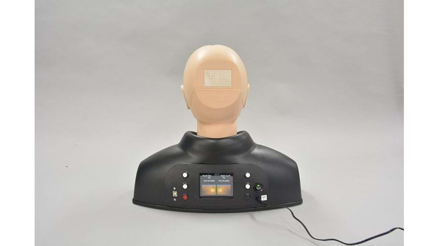
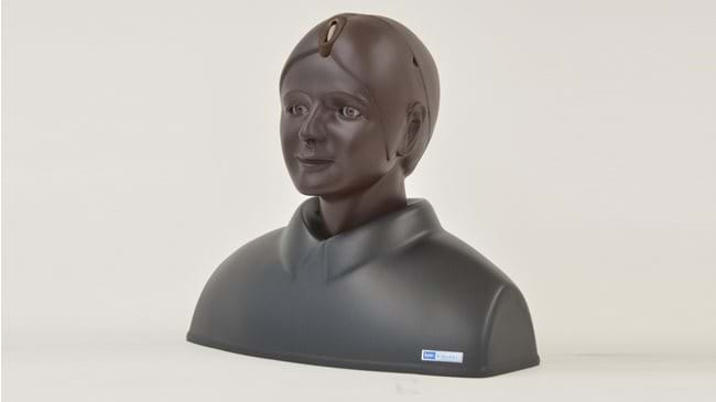
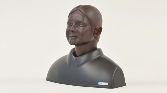
Digital Eye Examination Simulator (Light Skin Tone)
Kyoto Kagaku's Digital Eye Examination trainer builds upon it's popular standard Eye trainer, with upgraded components and a wider range of fundus images to diagnose.
This advanced simulator includes 40 fundus images, including 10 common diseases, which are pre-installed for ease of use. All the images are taken from actual clinical cases to give accurate examples of each disease.
What eye diseases can I identify using this simulator?
The following examples can be used for training:
- Normal eye-ground
- Hypertensive retinopathy: arteriolar vasoconstriction grade 3, arteriolosclerosis grade 1
- Haemorrhages and cotton wool spots, simple vein concealment
- Simple/background diabetic retinopathy: microaneurysm, hemorrhages and hard exudates
- Papilloedema (chronic phase)
- Papilloedema (acute phase)
- Glaucomatous optic atrophy: glaucomatous optic disc cupping and nerve fiber defect
- Retinal vein occlusion (acute phase): flame-shaped hemorrhage and cotton wool spots
- Retinal vein occlusion (after retinal laser photocoagulation)
- Toxoplasmosis: retinochoroiditis
- Age-related macular degeneration: macular exudates and subretinal hemorrhage
- 3 types of Enlarged optic disc recess
- Enlarged optic disc recess (Tilted disc)
- Enlarged optic disc recess (Infiltrate spot)
- 2 types of Drusen
- 2 types of Enlarged optic disc recess with drusen
- Macular Drusen
- Optic disc haemorrage
- Haemorrage
- 2 types of Chorioretinal atrophy
- 4 types of Vitreous clouding
- 2 types of Retinal nerve fiber layer defect
- Pigmentation
- Haemorrage with Vascular white sheath
- Photocoagulation spots
- 3 types of Epiretinal membrane
- Keratoconus
- Macular Degeneration
- Vitiligo
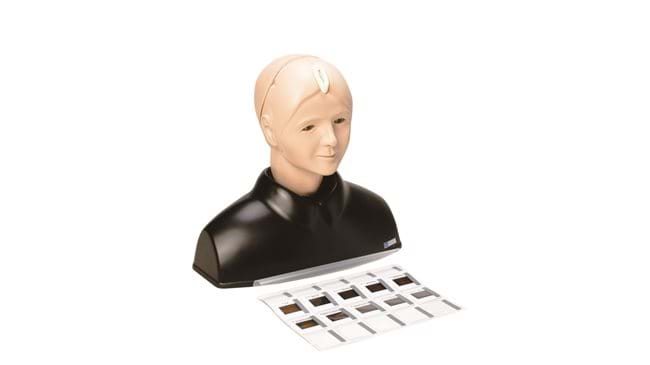
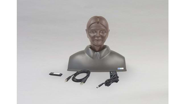
Note: It is also possible to add custom fundus images to the simulator via a USB connection.
Features
Overview
- Includes 40 pre-programmed fundus images
- Allows for custom image upload via USB connection
Realism
- Fundus images are from actual cases
Versatility
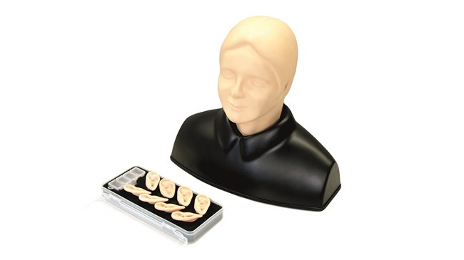
- No loose slides, easily switch between case images
- Compact all in on unit, easy to move during training
Safety
- Contains electronics, do not disassemble, or use near water sources
Anatomy
- Adult head and shoulders
Skills
Skills Gained
Identifying eye diseases, including:
- Hypertensive retinopathy
- Haemorrhages and cotton wool spots
- Simple/background diabetic retinopathy
- Papilloedema (chronic phase)
- Papilloedema (acute phase)
- Glaucomatous optic atrophy
- Retinal vein occlusion (acute phase)
- Retinal vein occlusion (after retinal laser photocoagulation)
- Toxoplasmosis: retinochoroiditis
- Age-related macular degeneration
- Enlarged optic disc recess
- Enlarged optic disc recess (Tilted disc and Infiltrate spot)
- Types of Drusen
- Enlarged optic disc recess with drusen
- Macular Drusen
- Optic disc haemorrage
- Chorioretinal atrophy
- Vitreous clouding
- Retinal nerve fiber layer defect
- Pigmentation
- Haemorrage with Vascular white sheath
- Photocoagulation spots
- Types of Epiretinal membrane
- Keratoconus
- Macular Degeneration
- Vitiligo
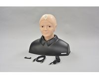
Contains
Check out our Ranges for specialised training sets
Browse our ranges