National Organisation of Nurse Practitioner Faculties, Nurse Practitioner Core Competencies Content
, 2017 Independent Practice Competencies p.14
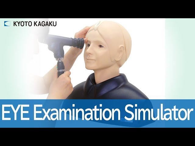
3.b Uses advanced health assessment skills to differentiate between normal, variations of normal and abnormal findings.
3.c Employs screening and diagnostic strategies in the development of diagnoses.