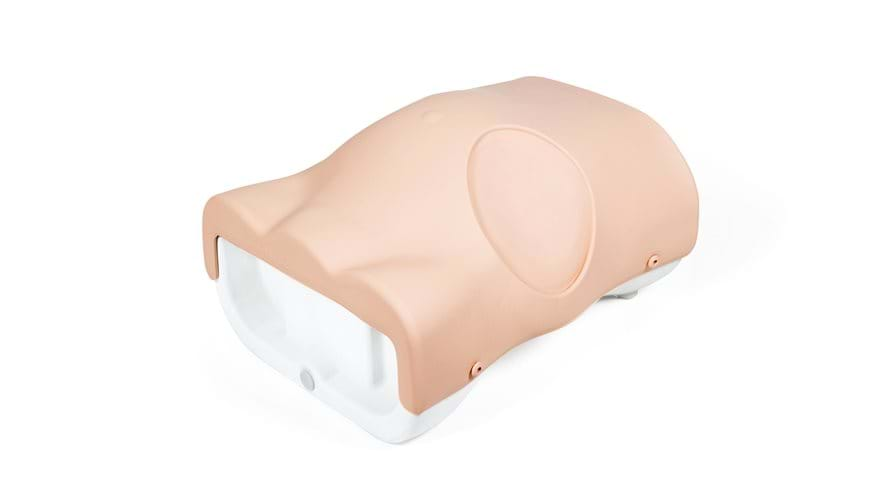


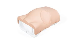

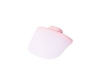
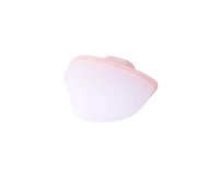
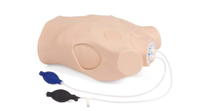
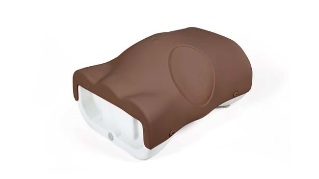
Paracentesis Trainer (Light Skin Tone)
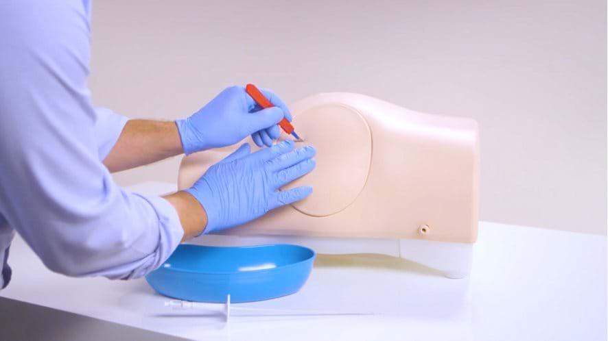
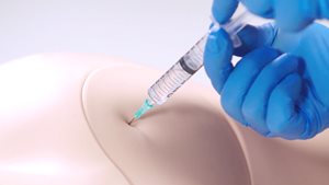
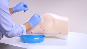
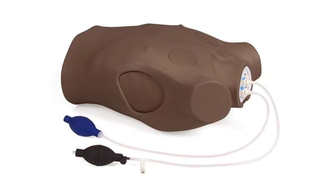
The Paracentesis Trainer offers dual functionality for diagnostic and therapeutic procedures. Palpation or ultrasound guided techniques can be used in the lower abdominal quadrants for the insertion of the needle and catheter while avoiding the liver, spleen and bowel.
Effective simulation training is essential, as paracentesis is a common procedure that’s practiced across a wide range of disciplines around the world.
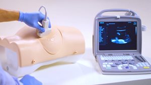
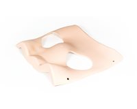
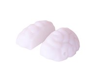
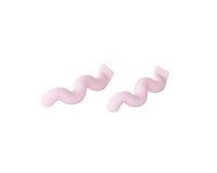
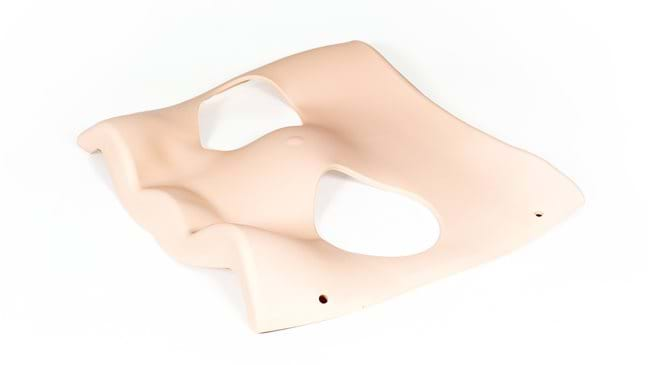
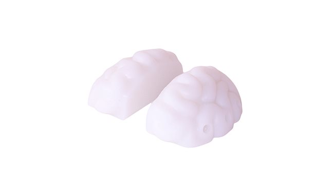
Echolucent pads and Echogenic organs inside the trainer give trainees the opportunity to become familiar with the internal anatomy under ultrasound, and recognizing how to safely insert a needle of catheter into the peritoneal cavity.
Overview
- Landmark or ultrasound techniques can be practiced (side by side)
- Internal echogenic anatomy to allow recognition of landmarks under ultrasound
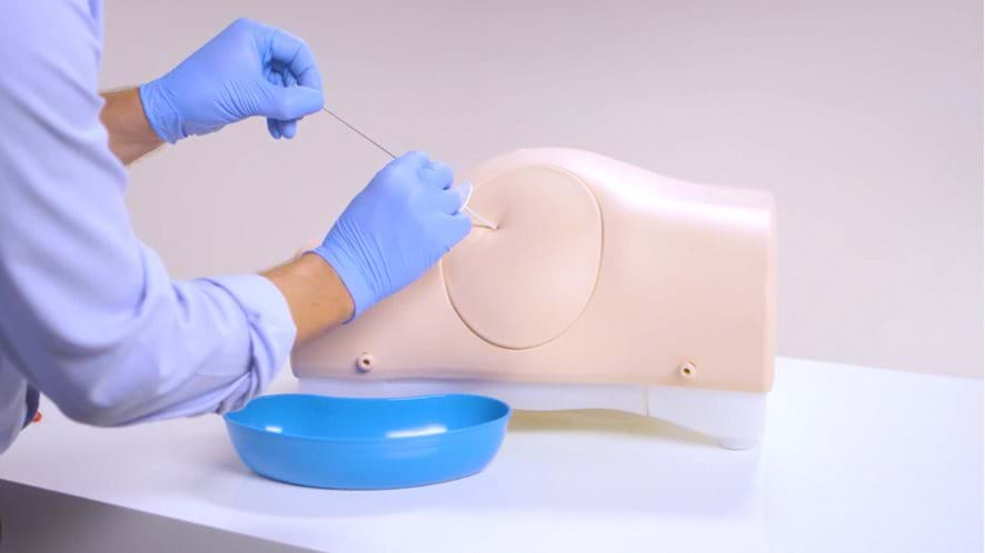
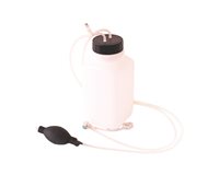
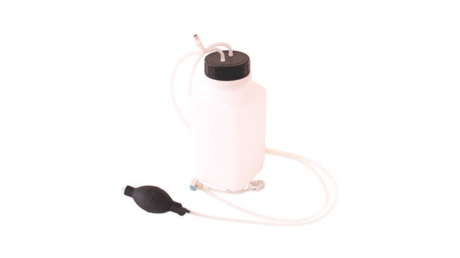
- Two 3.5 liter chambers can be filled with water for drainage practice
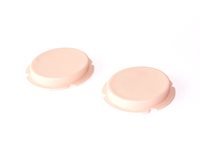
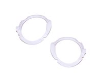
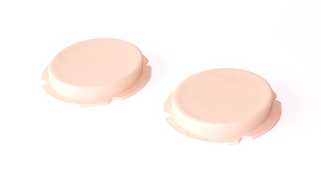
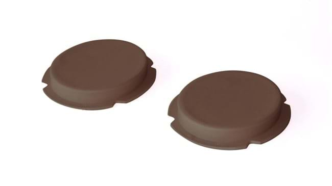
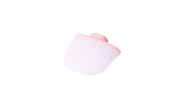
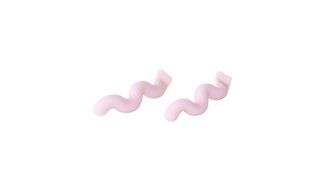
- Cost effective consumable pads
Realism
- Realistic tissue and needle response
Versatility
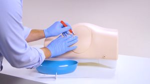
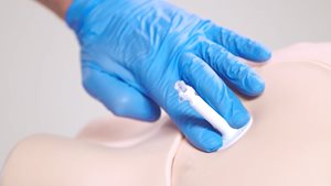
- Self sealing pads withstand up to 200 needle or up to 100 rocket catheter insertions
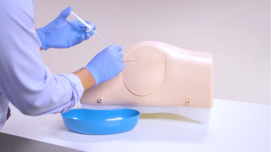
- Ability to insert and remove drain
- Positions both supine and lying on side
Cleaning
- Skin surface is washable using soap and water
Safety
- Latex free
Simulated Patient
- This product can be used with a simulated patient
Anatomy
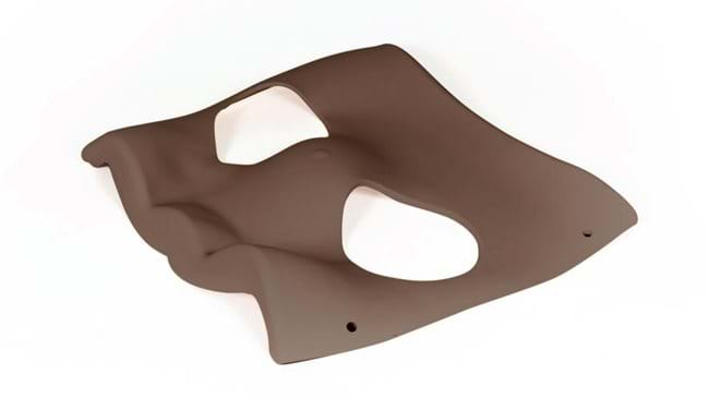
- Torso featuring bony landmarks and umbilicus
- Internal anatomy includes:
- Liver
- Spleen
- Bowel
- Floating Bowel
Skills Gained
- Familiarity with the abdominal regions and underlying anatomy
- Palpation of anatomical landmarks
- Identification of excess fluid
- Using ultrasound guidance, trainees can visualize the insertion site and check for vital organs beneath
- Insertion of needle into the peritoneal cavity for therapeutic or diagnostic purposes
- Professional-to-patient communication
Also supplied:
- Paracentesis Base Unit
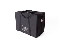
- Carry Case
References
Surgery
SURGICAL COUNCIL ON RESIDENT EDUCATION, Curriculum Outline for General Surgery 2017–2018 Operations/Procedures... Paracentesis
How much peritoneal fluid can I aspirate per training session?
Each of the two water cavities can hold up to 3.5l of “peritoneal” fluid at a time. However, using the large volume refill system, allows you to top up levels during training with minimal downtime.
NOTE: Take care when moving the model, with both cavities filled to capacity, the trainer’s weight can increase by around 7kg.
Can trainees practice injecting fluids into the abdomen?
Though it would be possible to inject fluids into the paracentesis pads, we do not recommend it. Introducing fluid or air to the pads can compromise the material inside, leading to a reduction in its lifespan. To practice the administering of anesthesia, we would recommend using an empty syringe.
Why does the Paracentesis Trainer not offer the option to perform midline procedures?
During the development stages of a product, we conduct market research with medical professionals in related fields. Following feedback on the Paracentesis Trainer, left lateral and right lateral approaches were deemed to be the most common in practice, and therefore the most important for this trainer.

Check out our Ranges for specialized training sets
Browse our ranges