
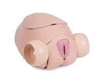
SFPT Mk 2 Uterus
The SFPT Uterus contains anatomical landmarks and pathologies for the practice of gynecological surgical procedures and techniques used within the surgical female pelvic trainer. This includes surgical skills to deal with:
- Salpingostomy
- Salpingectomy
- Cyctectomy
- Myomectomy
- Hysterectomy
- Oophorectomy
Compatible for use with harmonic scalpels and a range of surgical devices and instruments (See versatility). Included with the uterus are blood vessels that can be connected to a blood reservoir to simulate blood flow.
Overview
- Realistic bleeding when arteries are cut
- Anatomical pathologies and landmarks for realistic simulation
Realism
- Insufflation of the abdomen is represented
Versatility
For use with harmonic scalpels and other surgical instruments, including:
- Uterine manipulator – Rumi II
- Uterine sound – Hegar type
- Tenaculum (Cervical forceps)
- Speculums (Cusco, Sims, Grave)
Safety
- Contains Latex (Ovarian cyst which is enclosed in silicone)
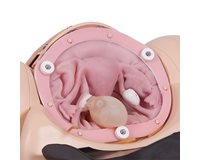
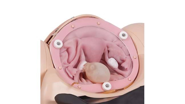
Anatomy
The uterus contains the following anatomical landmarks:
- Fallopian Tube
- Ovaries
- Broad Ligament
- Ureters
- Uterine Artery
- Ovarian Artery
- Pouch of Douglas
- Dunns Pouch
- Bowel and Sigmoid
- Ovarian Cyst
- Ectopic Pregnancy
- Fibroid
- Uterosacral Ligament
- Cardinal Ligament
Skills Gained
- Dissection down to and location of ureters
- Insertion and use of uterine manipulator
- Working with vagynecologicalogical instruments
- Identification of anatomical landmarks
- Practice and role-playing for gynecological perioperative teams
When used in conjunction with the Surgical Female Pelvic Trainer the skills that are developed include the following procedures:
- Salpingectomy
- Salpingostomy
- Myomectomy
- Cystectomy
- Hysterectomy
- Oophorectomy

Check out our Ranges for specialised training sets
Browse our Ranges