-
 New
New -

-

-
-
-

-
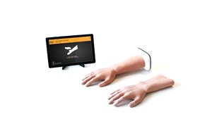

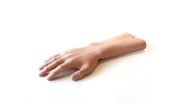
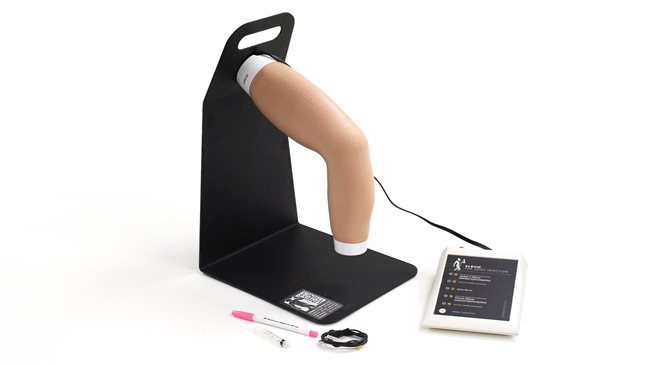
Hand & Wrist Injection Trainer (With Tablet, Light Skin Tone)
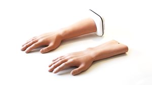
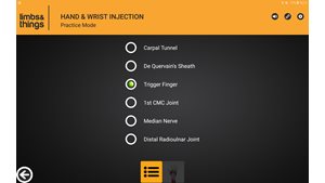
SuitSuitable for all levels of training, the Hand & Wrist Injection Trainer allows trainees and clinicians to practice and teach injections in five different locations within the hand and wrist.
An anatomically correct representation of an adult hand and partial forearm with important landmarks and underlying structures, the palpation-guided trainer allows for injections into the:
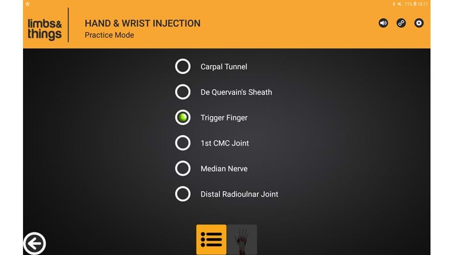
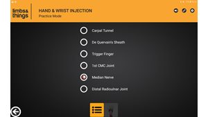
• Carpal Tunnel
• De Quervain’s Sheath
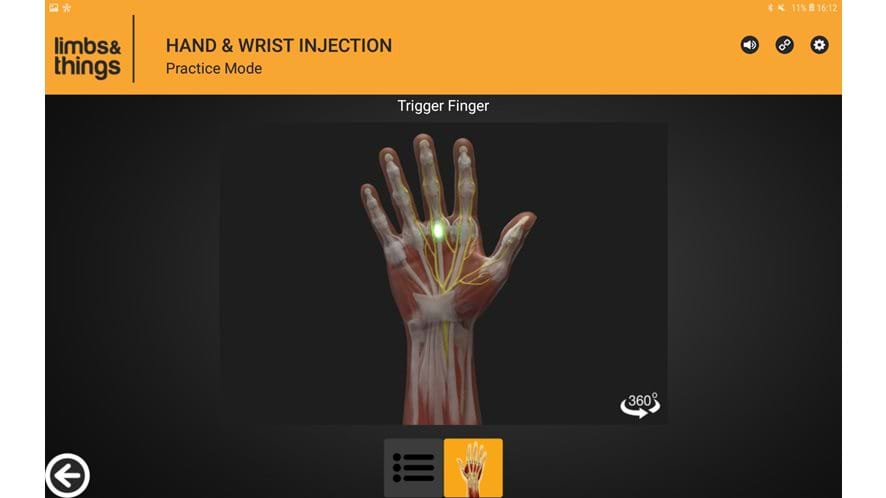
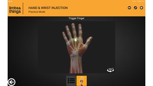
• Trigger Finger
• First Carpometacarpal Joint
• Distal Radioulnar Joint
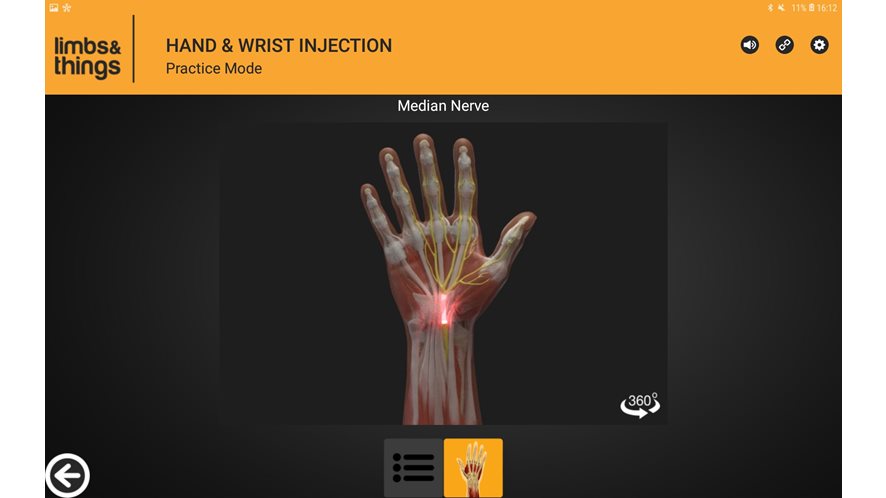
The app, coupled with innovative sensory technology, provides learners with wireless, real-time feedback from the procedure. An Interactive Anatomy element allows users to rotate the detailed model anatomy 360 degrees so each injection site can be viewed and analysed from a number of angles.
able for all levels of training, the NEW Hand & Wrist Injection Trainer allows trainees and clinicians to practice and teach injections in five different locations within the hand and wrist. An anatomically correct representation of a man’s hand with important landmarks and underlying structures, the palpation-guided trainer allows for injections into the Carpal Tunnel, De Quervain’s Sheath, Trigger Finger, First Carpometacarpal Joint, and the Distal Radioulnar Joint, whilst the presence of the Median Nerve adds another level of realism and complexity.
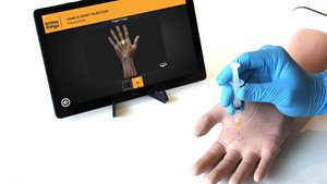
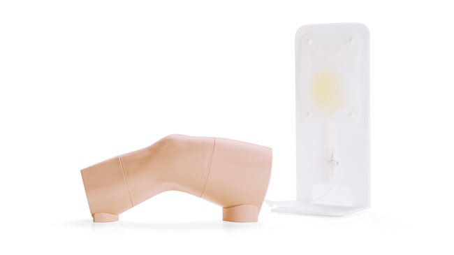
The trainer comes equipped with an Android tablet, which works in unison with our newly developed app. The app, coupled with innovative sensory technology, provides learners with wireless, real-time feedback from the procedure. An Interactive Anatomy element allows users to rotate the detailed model anatomy 360 degrees so each injection site can be viewed and analysed from a number of angles.
A mobile android tablet is provided which is guaranteed compatible with the hand & wrist injection trainer. The application is pre-installed on the tablet provided. If you already have a compatible tablet then a hand & wrist injection trainer can be purchased without a tablet.
Overview
• Facilitates injection in five key sites of the hand and wrist
• Presence of the Median Nerve allows for negative feedback
• App provides interactive real-time feedback
• Each site can withstand extensive repeated injections
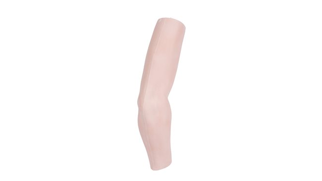
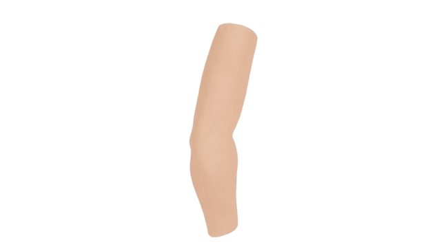
• Skin can be easily removed and changed (see skin replacement)
• Interactive model anatomy
Realism
- Interactive model anatomy
- Anatomically accurate male adult
- Includes important ligament and skeletal structures (See Anatomy)
Versatility
- Lightweight and easily portable
- Can be used alongside a simulated patient for communication training
- App can be set to English UK, English USA, French, Mandarin, Spanish and German
Cleaning
- Can be cleaned by wiping with soap and water
Safety
- Fluid should not be injected into any of the sites
- Take care when cleaning trainer as electronics are present
- This trainer is latex free
Anatomy
Injection Sites
- Carpal Tunnel
- De Quervain’s Sheath
- Trigger Finger
- First Carpometacarpal Joint
- Distal Radioulnar Joint
- Median Nerve
- Trigger finger nodule
Internal Product Anatomy:
- Palmaris Longus
- Flexor Carpi Radialis
- Extensor Digitorum Group
- Extensor Digiti Minimi
- Abductor Pollicus Longus
- Extensor Pollicus Brevis
- Extensor Pollicus Longus
Bone structures:
- Radial Styloid
- Ulnar Styloid
- Trapezius and Scaphoid
- First Carpometacarpal Joint space
- Distal Radioulnar Joint space
- Lister’s tubercle
Skills Gained
- Model also includes technology that ensures trainees learn to avoid the Median Nerve
- Patient positioning and management
- Communication skills when used with a simulated patient
- Identification of anatomical landmarks
Injecting into the:
- Carpal Tunnel
- De Quervain’s Sheath
- Trigger Finger
- 1st Carpometacarpal Joint
- Distal Radioulnar Joint
-

-

-

Android tablet
-
AA batteries (x2)
-
Sharps Bin
References
Undergraduate
Recommendations for Clinical Skills Curricula for Undergraduate Medical Education, Association of American Medical Colleges, 2008, Appendix B; Clinical Testing and Procedural Skills, p.26 Perform and describe systematic examination of major joints including inspection, palpation, range of motion, and identification of important anatomical landmarks Recommendations for Clinical Skills Curricula for Undergraduate Medical Education, Association of American Medical Colleges, 2005, Appendix B; Clinical Testing and Procedural Skills, p.23 Testing & Procedure Skills: Joint Aspiration Technique; Joint Fluid Examination” Procedural skills: What’s taught in medical school, what ought to be? SR Turner, J Hanson, CJ de Gara, Non-AAMC Recommended: Joint aspiration - % of Schools That Teach - 88%
Family Medicine Residency
Graduate Medical Education in Pain Medicine
Graduate Medical Education in Physical Medicine and Rehabilitation
Graduate Medical Education in Orthopaedic Surgery
Rheumatology Fellows
Graduate Medical Education in Musculoskeletal Radiology
Nurse Practitioner (NP) and Physician Assistant (PA) Specialising in Rheumatology
Physician Assistant (PA) Specialising in Orthopaedics
Can I use this hand & wrist injection simulator for self-directed learning?
Yes, the hand & wrist simulator has an accompanying app which allows trainees to receive instant positive & negative feedback from the model depending on whether the correct injection site is hit.
What is the expected battery life for the Hand & Wrist Injection Trainer?
Operated by 2 x AA batteries, the trainer has undergone laboratory testing, and we’ve shown that the batteries can last for over 500 hours of injection practice.
What are the requirements to use the app on my own device?
The trainer works over Bluetooth, so a Bluetooth enabled Android™ tablet or phone is required for training. This allows the needles and device to be completely wireless, ensuring maximum realism when injecting sites
Can I practise different injection techniques on this model?
The Hand & Wrist trainer is anatomically accurate, meaning a wide range of techniques and approaches can be taken with the model to ensure trainees can hit a gold standard of training.

Check out our Ranges for specialized training sets
Browse our ranges