


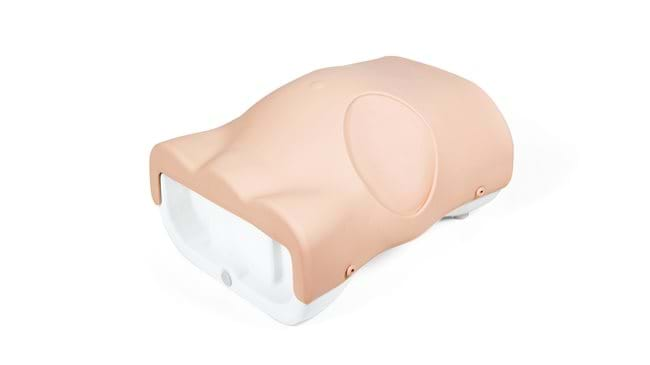
Combined Ultrasound Guided Thoracentesis/ Pericardiocentesis
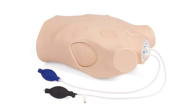
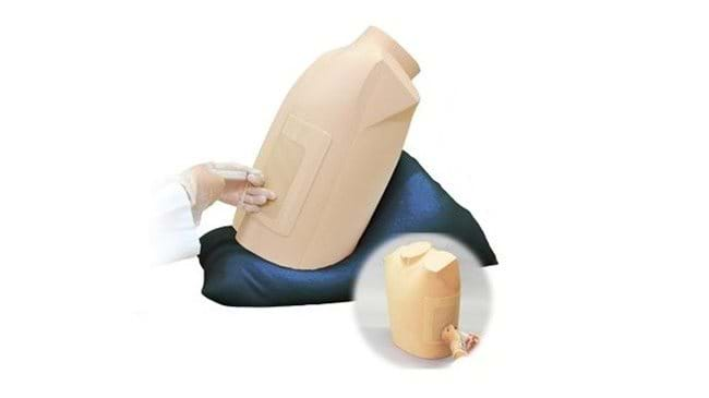
This ultrasound compatible model is a multifaceted trainer that facilitates training in thoracentesis and pericardiocentesis. The combined model allows trainees to use the clear ultrasound image provided to accurately perform both procedures.
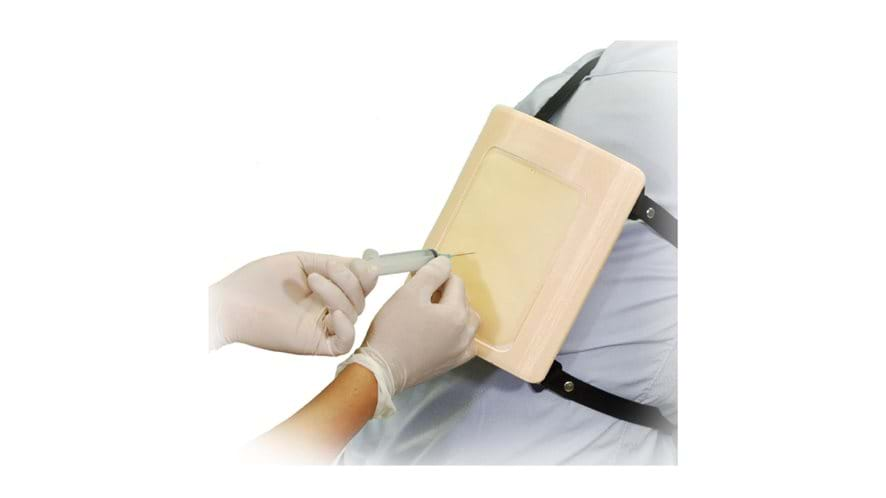
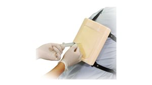
The Thoracentesis Trainer accommodates mid-scapular line access and mid-axillary line access, whilst the strap-on units allow for the use of a simulated patient in a training scenario.
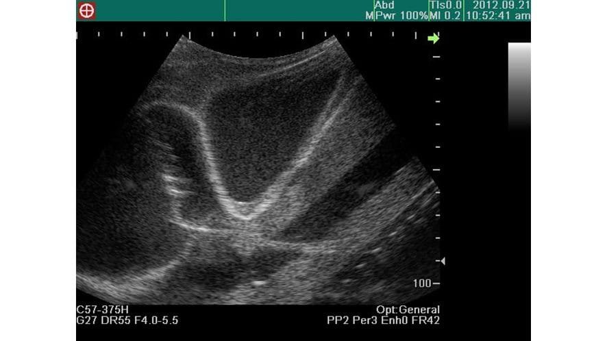
The ability to pierce the pericardial sac using ultrasound guidance provides learners with the platform to repeatedly practice patient safety during the pericardiocentesis procedure.
Overview
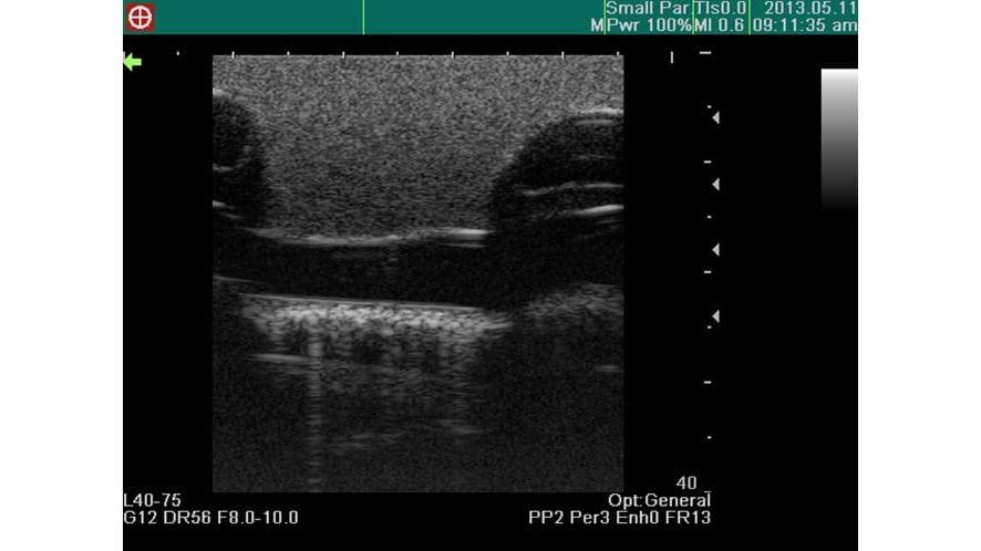
- Excellent ultrasound image
- Anatomically correct puncture sites reproduce realistic needle-top resistance and sensation
- Clear feedback on successful or unsuccessful procedures
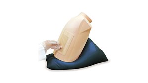
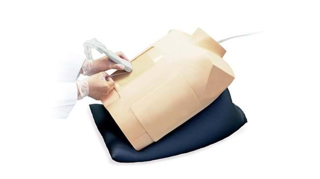
- Strap-on units enhance the learning of patient positioning and professional-to-patient communication
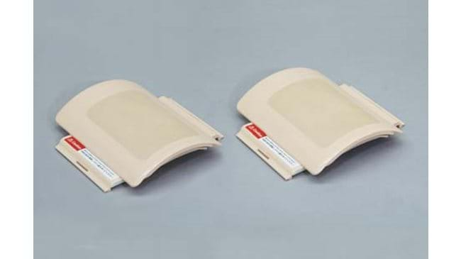
- Durable and replaceable puncture pads
Realism
Thoracentesis
- Palpable ribs
Pericardiocentesis
- Realistic needle tip feeling when the needle pierces the pericardial sac
- Confirmation of ventricles, ribs, pericardium, liver and main artery under ultrasound scanning
Versatility
- Two sites for access: right mid-scapular line and left mid-axillary line on the Thoracentesis Trainer
- Volume of pleural effusion can be controlled on the Thoracentesis Trainer to set different levels of challenges
- The upper torso manikin can be set in different positions to learn patient positioning for both procedures
Anatomy
- Left Xiphisternal junction (Larrey’s point)
- Anatomical parts include ribs, pleura, soft tissue and diaphragm
- Mid-axillary line: 6-9th ribs
- Mid-scapular line: 8-11th ribs
Skills Gained
Thoracentesis
- Recognition of anatomical landmarks using ultrasound
- Assessment of level and volume of pleural effusion
- Determination of insertion site
- Needle insertion and collection of fluid
- Patient positioning
Pericardiocentesis
- Ability to practice both the subxiphoid approach and parasternal approach
- Visualization of pericardial fluid using ultrasound scanning
- Landmark palpation
- Identification of ‘Larrey’s point’ for needle insertion
- Needle insertion to the pericardial space
- Aspiration of pericardial effusion
- Patient positioning
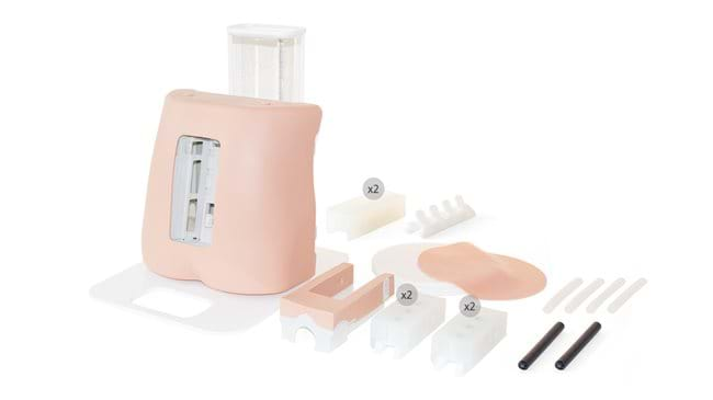
-

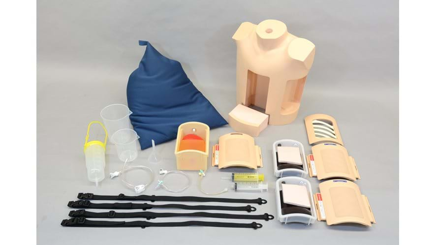
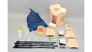
1 Adult Chest Model
-
1 Mid-Axillary Line Unit
-
1 Mid-scapular Line Unit
-
1 Pericardiocentesis Unit
-
1 Pillow for Positioning
-
1 Explanation Model for Thoracentesis
-
1 Instruction Manual
References
Intensive Care Medicine
ICM Curriculum: Supporting Excellence for a CCT in Intensive Care Medicine, The Faculty of Intensive Care Medicine, 2021. Drainage of fluid.
Trauma and Orthopaedic

Check out our Ranges for specialised training sets
Browse our ranges